
In a remarkable breakthrough that could transform cancer surgery, Canadian researchers have discovered that the brain emits a subtle natural glow that can help distinguish cancerous cells from healthy tissue. This innovative approach promises to give surgeons a powerful new tool in the fight against aggressive brain cancers.
The Science Behind the Glow
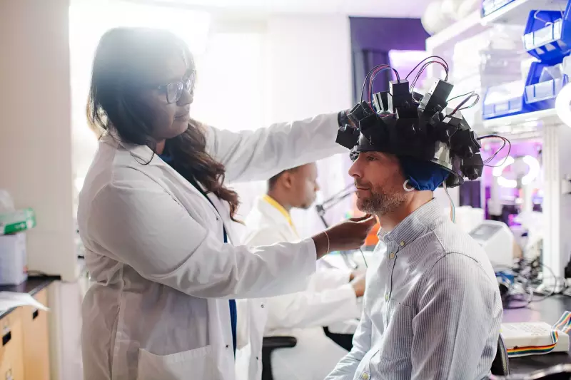
Researchers at the University of Waterloo and Western University have uncovered that brain tissue naturally fluoresces under specific lighting conditions. What's truly revolutionary is that cancerous cells glow differently than healthy brain cells, creating a visible distinction that surgeons can use during operations.
Dr. Parsin Haji Reza, the principal investigator and professor of systems design engineering at Waterloo, explains the significance: "What we've discovered is that the brain has its own inherent glow when we apply specific light wavelengths. More importantly, cancer cells and healthy cells glow with different intensities and patterns, giving us a clear visual marker."

Transforming Brain Cancer Surgery
The technology, called photo-histology, represents a major advancement over current methods. Traditionally, surgeons must remove tissue samples and wait for pathology lab results during surgery—a process that can take 30-40 minutes and doesn't always provide clear boundaries between cancerous and healthy tissue.
With this new approach, surgeons could potentially see cancer boundaries in real-time, allowing for more precise removal of tumors while preserving crucial healthy brain tissue. This is particularly critical for aggressive cancers like glioblastoma, where complete tumor removal significantly impacts patient outcomes.
How the Technology Works
The research team uses specialized microscopes that apply specific wavelengths of light to brain tissue. The natural fluorescence that results provides immediate visual feedback about tissue health. The method has shown remarkable accuracy in early testing, correctly identifying cancerous tissue with high precision.
Unlike artificial dyes or contrast agents that require injection into the body, this technique relies on the brain's natural properties, making it safer and more accessible for widespread surgical use.
Future Applications and Impact
While initially focused on brain cancer, researchers believe this technology could eventually help with other types of cancer surgery where clear boundaries between healthy and diseased tissue are crucial. The team continues to refine the technology and hopes to begin clinical trials in the near future.
This Canadian-led innovation represents a significant step forward in the global fight against cancer, potentially giving surgeons the ability to see what was previously invisible and saving countless lives through more precise surgical interventions.